


Advancing neurosurgical outcomes with real-time diagnostic imaging
The first moveable, intraoperative MRI system in the Washington region is at MedStar Georgetown University Hospital.
The Intraoperative MRI System (IMRIS) at MedStar Georgetown University Hospital uses MRI technology to digitally map a patient’s brain, head, and neck during brain surgery. IMRIS is the only iMRI system in the world that travels on ceiling-mounted rails between diagnostic rooms and operating rooms. It brings MRI directly to the patient and provides real-time imaging during surgery to neurosurgeons and their surgical team. The system allows neurosurgeons to precisely remove brain tumors and accurately place electrodes during deep brain stimulation surgeries all while reducing the need for post-op imaging and additional surgeries. This technology allows surgeons to make informed decisions during the operation, leading to better outcomes for patients.
For brain cancer patients, IMRIS helps ensure that the entire tumor is removed during surgery, offering numerous benefits to the patient, such as:
- Fewer repeat surgeries
- Less post-operative imaging
- Better outcomes and protection of surrounding tissues and brain function
- Peace of mind for patients and their families
The addition of the IMRIS technology also improves the treatment of several neurological, spinal, and cerebrovascular disorders. IMRIS is most frequently used in procedures related to:
- Brain tumors
- Epilepsy
- Deep brain stimulator electrode insertion
- Essential tremor
- Parkinson’s disease
- Other movement disorders
IMRIS is just one of the many breakthrough technologies the world-class neurosurgery team at MedStar Health offers.
IMRIS video
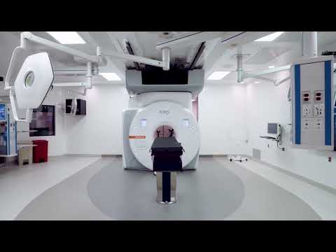

IMRIS at the Verstandig Pavilion at MedStar Georgetown University Hospital
The impact of IMRIS on patient outcomes
- Surgeons achieved a 28-point increase in the percentage of patients with gross/total resection when iMRI was used.
- In 49% of all cases, the surgeon modified the procedure based on the findings of iMRI – information that would not have been available until post-procedure.
- In 52% of glioma cases, the surgeon resected additional tumor that was identified using iMRI.